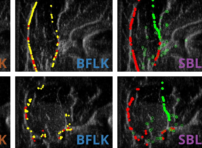
 Various optical flow methods show promise in tracking the fascial contour of the brachioradialis muscle during isometric contraction.
Muscle Deformation Correlates with Output Force during Isometric Contraction
 Various optical flow methods show promise in tracking the fascial contour of the brachioradialis muscle during isometric contraction.
Muscle Deformation Correlates with Output Force during Isometric Contraction
Abstract
Despite the utility of musculoskeletal dynamics modeling, there exists no safe, noninvasive method of measuring in vivo muscle output force in real time. In this paper, we demonstrate that muscle deformation constitutes a promising, yet unexplored class of signals from which to infer such forces. Through a preliminary case study of the elbow joint, in which we examine simultaneous flexion force, surface electromyography (sEMG), and ultrasound imaging data during isometric contraction, we provide evidence that even simple measures of deformation (including cross-sectional area and thickness variation in the brachioradialis muscle) correlate well with elbow output torque to an extent comparable with standard sEMG activation measures. We then show that these simple signals, as well as the overall brachioradialis contour, can be tracked over time via optical flow techniques, enabling the use of these signals in real-time applications (e.g., assistive device control), as well as larger-scale study of deformation signals without necessitating manual annotation. To enable such future work, all modeling and tracking software described in this paper, as well as all raw and processed data, have been made available on SimTK as part of the OpenArm project (https://simtk.org/projects/openarm) for general research use.
